
Fil:Viral luciferase expression in a mouse tumour.jpg
Det finns ingen version med högre upplösning.
Viral_luciferase_expression_in_a_mouse_tumour.jpg (229 × 346 pixlar, filstorlek: 54 kbyte, MIME-typ: image/jpeg)
| Denna fil tillhandahålls av Wikimedia Commons. Informationen nedan är kopierad från dess filbeskrivningssida. |
Sammanfattning
| Beskrivning |
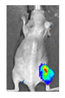
English: A 'nude' (hairless) mouse bearing a tumour in the right hind leg has been treated intra-tumourally with vaccinia virus encoding luciferase. Viral infection of the tumour is visible by the bio-luminescence produced by luciferase. (False coloring is used to demonstrate intensity). Citation: Haddad D, Chen C-H, Carlin S, Silberhumer G, Chen NG, et al. (2012) Imaging Characteristics, Tissue Distribution, and Spread of a Novel Oncolytic Vaccinia Virus Carrying the Human Sodium Iodide Symporter. PLoS ONE 7(8): e41647. doi:10.1371/journal.pone.0041647 |
| Datum | |
| Källa | http://www.plosone.org/article/info%3Adoi%2F10.1371%2Fjournal.pone.0041647 |
| Skapare | Haddad D, Chen C-H, Carlin S, Silberhumer G, Chen NG, et al. |
| Tillstånd (Återanvändning av denna fil) |
http://www.plosone.org/static/license;jsessionid=D9B1E557E36EDD0EDB521F6783647855 |
Denna fil har gjorts tillgänglig under licensen Creative Commons Erkännande 1.0 Generisk
- Du är fri:
- att dela – att kopiera, distribuera och sända verket
- att remixa – att skapa bearbetningar
- På följande villkor:
- erkännande – Du måste ge lämpligt erkännande, ange en länk till licensen och indikera om ändringar har gjorts. Du får göra det på ett lämpligt sätt, men inte på ett sätt som antyder att licensgivaren stödjer dig eller din användning.
Licensiering
Denna fil har gjorts tillgänglig under licensen Creative Commons Erkännande 2.5 Generisk
- Du är fri:
- att dela – att kopiera, distribuera och sända verket
- att remixa – att skapa bearbetningar
- På följande villkor:
- erkännande – Du måste ge lämpligt erkännande, ange en länk till licensen och indikera om ändringar har gjorts. Du får göra det på ett lämpligt sätt, men inte på ett sätt som antyder att licensgivaren stödjer dig eller din användning.
Filhistorik
Klicka på ett datum/klockslag för att se filen som den såg ut då.
| Datum/Tid | Miniatyrbild | Dimensioner | Användare | Kommentar | |
|---|---|---|---|---|---|
| nuvarande | 30 maj 2013 kl. 19.43 | | 229 × 346 (54 kbyte) | Viraltonic | {{subst:Upload marker added by en.wp UW}} {{Information |Description = {{en|A 'nude' (hairless) mouse bearing a tumour in the right hind leg has been treated intra-tumourally with vaccinia virus encoding luciferase. Viral infection of the tumour is vis... |
Filanvändning
Följande sida använder den här filen:
Global filanvändning
Följande andra wikier använder denna fil:
- Användande på ar.wikipedia.org
- Användande på en.wikipedia.org
- Användande på ja.wikipedia.org